Pseudomonas aeruginosa PAO1, PA4261 (rplW)
Cytoplasmic
Cytoplasmic Membrane
Periplasmic
Outer Membrane
Extracellular
Unknown
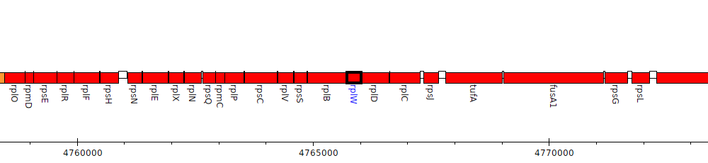
Gene Feature Overview
| Strain |
Pseudomonas aeruginosa PAO1 (Stover et al., 2000)
GCF_000006765.1|latest |
| Locus Tag |
PA4261
|
| Name |
rplW
|
| Replicon | chromosome |
| Genomic location | 4765713 - 4766012 (- strand) |
Cross-References
| RefSeq | NP_252951.1 |
| GI | 15599457 |
| Affymetrix | PA4261_rplW_at |
| Entrez | 881753 |
| GenBank | AAG07649.1 |
| INSDC | AAG07649.1 |
| NCBI Locus Tag | PA4261 |
| protein_id(GenBank) | gb|AAG07649.1|AE004841_27|gnl|PseudoCAP|PA4261 |
| TIGR | NTL03PA04262 |
| UniParc | UPI00000C5C8B |
| UniProtKB Acc | Q9HWD7 |
| UniProtKB ID | RL23_PSEAE |
| UniRef100 | UniRef100_A6UZJ0 |
| UniRef50 | UniRef50_B8H4D6 |
| UniRef90 | UniRef90_A6UZJ0 |
Product
| Feature Type | CDS |
| Coding Frame | 1 |
| Product Name |
50S ribosomal protein L23
|
| Synonyms | |
| Evidence for Translation |
Identified using nanoflow high-pressure liquid chromatography (HPLC) in conjunction with microelectrospray ionization on LTQ XL mass spectrometer (PMID:24291602).
|
| Charge (pH 7) | 9.22 |
| Kyte-Doolittle Hydrophobicity Value | -0.421 |
| Molecular Weight (kDa) | 10.9 |
| Isoelectric Point (pI) | 10.81 |
Subcellular localization
| Individual Mappings | |
| Additional evidence for subcellular localization |
PDB 3D Structures
| Accession | Header | Accession Date | Compound | Source | Resolution | Method | Percent Identity |
| 6SPF | RIBOSOME | 09/01/19 | Pseudomonas aeruginosa 70s ribosome from an aminoglycoside resistant clinical isolate | Pseudomonas aeruginosa | 2.89 | ELECTRON MICROSCOPY | 100.0 |
| 7UNR | RIBOSOME | 04/11/22 | Pseudomonas aeruginosa 70S ribosome initiation complex bound to compact IF2-GDP (composite structure I-A) | Escherichia coli; Pseudomonas aeruginosa PAO1; SYNTHETIC CONSTRUCT | 2.9 | ELECTRON MICROSCOPY | 100.0 |
| 7UNW | RIBOSOME | 04/11/22 | Pseudomonas aeruginosa 70S ribosome initiation complex bound to IF2-GDPCP (structure II-B) | Escherichia coli; Pseudomonas aeruginosa PAO1; SYNTHETIC CONSTRUCT | 2.6 | ELECTRON MICROSCOPY | 100.0 |
| 7UNV | RIBOSOME | 04/11/22 | Pseudomonas aeruginosa 70S ribosome initiation complex bound to IF2-GDPCP (structure II-A) | Escherichia coli; Pseudomonas aeruginosa PAO1; SYNTHETIC CONSTRUCT | 2.7 | ELECTRON MICROSCOPY | 100.0 |
| 7UNU | RIBOSOME | 04/11/22 | Pseudomonas aeruginosa 70S ribosome initiation complex bound to compact IF2-GDP (composite structure I-B) | Escherichia coli; Pseudomonas aeruginosa PAO1; SYNTHETIC CONSTRUCT | 2.9 | ELECTRON MICROSCOPY | 100.0 |
| 6SPG | RIBOSOME | 09/01/19 | Pseudomonas aeruginosa 70s ribosome from a clinical isolate | Pseudomonas aeruginosa | 3.34 | ELECTRON MICROSCOPY | 100.0 |
| 6SPD | RIBOSOME | 09/01/19 | Pseudomonas aeruginosa 50s ribosome from a clinical isolate | Pseudomonas aeruginosa | 3.28 | ELECTRON MICROSCOPY | 100.0 |
| 6SPB | RIBOSOME | 09/01/19 | Pseudomonas aeruginosa 50s ribosome from a clinical isolate with a mutation in uL6 | Pseudomonas aeruginosa | 2.82 | ELECTRON MICROSCOPY | 97.9 |
Pathogen Association Analysis
| Results |
Common
Found in both pathogen and nonpathogenic strains
Hits to this gene were found in 602 genera
|
Orthologs/Comparative Genomics
| Pseudomonas Ortholog Database | View orthologs at Pseudomonas Ortholog Database |
| Pseudomonas Ortholog Group |
POG001449 (535 members) |
| Putative Inparalogs | None Found |
Interactions
| STRING database | Search for predicted protein-protein interactions using:
Search term: PA4261
Search term: rplW
Search term: 50S ribosomal protein L23
|
Human Homologs
References
|
Primary structure of protein L23 from the Escherichia coli ribosome.
Wittmann-Liebold B, Greuer B
FEBS Lett 1979 Dec 1;108(1):69-74
PubMed ID: 391594
|
|
Structure of the Escherichia coli S10 ribosomal protein operon.
Zurawski G, Zurawski SM
Nucleic Acids Res 1985 Jun 25;13(12):4521-6
PubMed ID: 3892488
|
|
Incorporation of dinitrophenyl protein L23 into totally reconstituted Escherichia coli 50 S ribosomal subunits and its localization at two sites by immune electron microscopy.
Montesano-Roditis L, Glitz DG, Perrault AR, Cooperman BS
J Biol Chem 1997 Mar 28;272(13):8695-703
PubMed ID: 9079702
|