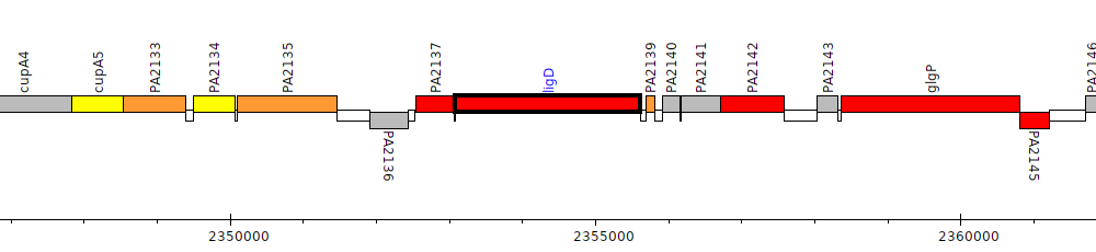
Pseudomonas aeruginosa PAO1, PA2138 (ligD)
Cytoplasmic
Cytoplasmic Membrane
Periplasmic
Outer Membrane
Extracellular
Unknown
Gene Feature Overview
| Strain |
Pseudomonas aeruginosa PAO1 (Stover et al., 2000)
GCF_000006765.1|latest |
| Locus Tag |
PA2138
|
| Name |
ligD
|
| Replicon | chromosome |
| Genomic location | 2353086 - 2355608 (+ strand) |
| Transposon Mutants | 2 transposon mutants in PAO1 |
| Transposon Mutants in orthologs | 1 transposon mutants in orthologs |
Cross-References
| RefSeq | NP_250828.1 |
| GI | 15597334 |
| Affymetrix | PA2138_at |
| Entrez | 880451 |
| GenBank | AAG05526.1 |
| INSDC | AAG05526.1 |
| NCBI Locus Tag | PA2138 |
| protein_id(GenBank) | gb|AAG05526.1|AE004641_5|gnl|PseudoCAP|PA2138 |
| TIGR | NTL03PA02138 |
| UniParc | UPI00000C55C8 |
| UniProtKB Acc | Q9I1X7 |
| UniProtKB ID | LIGD_PSEAE |
| UniRef100 | UniRef100_Q9I1X7 |
| UniRef50 | UniRef50_Q9I1X7 |
| UniRef90 | UniRef90_Q9I1X7 |
Product
| Feature Type | CDS |
| Coding Frame | 1 |
| Product Name |
Multifunctional non-homologous end joining protein LigD
|
| Synonyms |
probable ATP-dependent DNA ligase |
| Evidence for Translation | |
| Charge (pH 7) | 18.97 |
| Kyte-Doolittle Hydrophobicity Value | -0.530 |
| Molecular Weight (kDa) | 94.0 |
| Isoelectric Point (pI) | 9.72 |
Subcellular localization
| Individual Mappings | |
| Additional evidence for subcellular localization |
PDB 3D Structures
| Accession | Header | Accession Date | Compound | Source | Resolution | Method | Percent Identity |
| 3N9B | LIGASE | 05/28/10 | Crystal Structure of the P. aeruginosa LigD phosphoesterase domain | Pseudomonas aeruginosa | 1.92 | X-RAY DIFFRACTION | 100.0 |
| 2FAO | HYDROLASE/TRANSFERASE | 12/07/05 | Crystal Structure of Pseudomonas aeruginosa LigD polymerase domain | Pseudomonas aeruginosa | 1.5 | X-RAY DIFFRACTION | 100.0 |
| 2FAR | Hydrolase/Transferase | 12/07/05 | Crystal Structure of Pseudomonas aeruginosa LigD polymerase domain with dATP and Manganese | Pseudomonas aeruginosa | 1.9 | X-RAY DIFFRACTION | 100.0 |
| 2FAQ | HYDROLASE/TRANSFERASE | 12/07/05 | Crystal Structure of Pseudomonas aeruginosa LigD polymerase domain with ATP and Manganese | Pseudomonas aeruginosa | 1.9 | X-RAY DIFFRACTION | 100.0 |
| 2LJ6 | DNA BINDING PROTEIN | 09/06/11 | Solution Structure and DNA-binding Properties of the Phosphoesterase Domain of DNA Ligase D | Pseudomonas aeruginosa | NOT | SOLUTION NMR | 100.0 |
| 3N9D | LIGASE | 05/28/10 | Monoclinic Structure of P. aeruginosa LigD phosphoesterase domain | Pseudomonas aeruginosa | 2.3 | X-RAY DIFFRACTION | 100.0 |
Pathogen Association Analysis
| Results |
Common
Found in both pathogen and nonpathogenic strains
Hits to this gene were found in 290 genera
|
Orthologs/Comparative Genomics
| Pseudomonas Ortholog Database | View orthologs at Pseudomonas Ortholog Database |
| Pseudomonas Ortholog Group |
POG003336 (490 members) |
| Putative Inparalogs | None Found |
Interactions
| STRING database | Search for predicted protein-protein interactions using:
Search term: PA2138
Search term: ligD
|
Human Homologs
References
|
Characterization of an ATP-dependent DNA ligase encoded by Haemophilus influenzae.
Cheng C, Shuman S
Nucleic Acids Res 1997 Apr 1;25(7):1369-74
PubMed ID: 9060431
|
|
Gap filling activities of Pseudomonas DNA ligase D (LigD) polymerase and functional interactions of LigD with the DNA end-binding Ku protein.
Zhu H, Shuman S
J Biol Chem 2010 Feb 12;285(7):4815-25
PubMed ID: 20018881
|